

Gallery#
Reconstructions made with TIGRE, mostly from real scans, across medical, synchrotron, micro-CT, neutron and proton imaging. For papers that use TIGRE in each field, see Papers using TIGRE.
Medical cone-beam CT#
A RANDO head phantom, which represents an adult head, scanned at The Christie Hospital in Manchester with 360 equally spaced projections over a full rotation. The data have mechanical offsets and high noise, from head CBCT settings with low X-ray intensity.
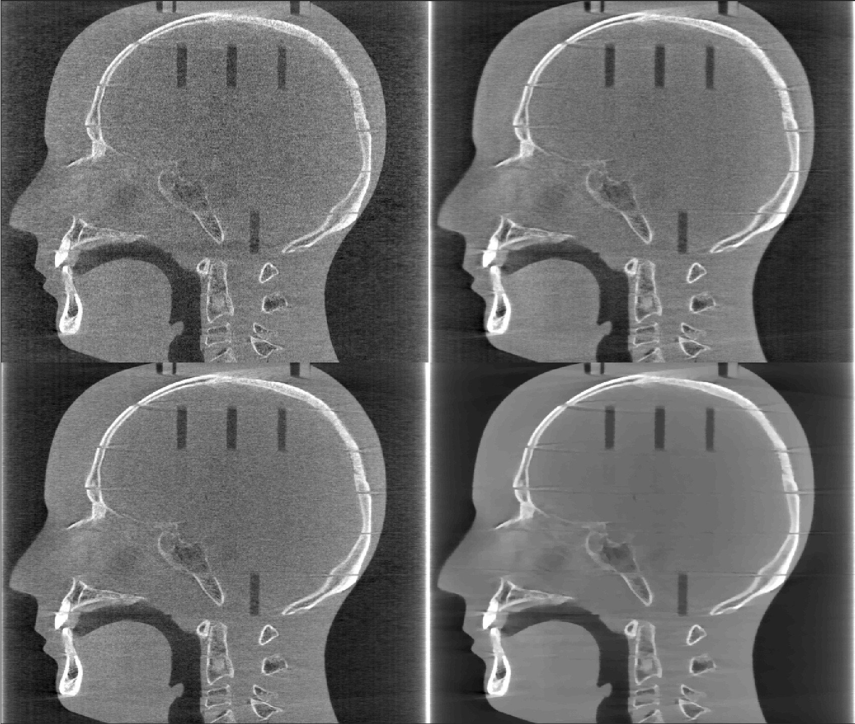
RANDO head from 360 projections. Left to right, top to bottom: FDK, CGLS, OS-SART and OS-ASD-POCS. Display window [0, 0.05].#

The same scan with only 90 projections, same algorithms and display window.#
Thanks to The Christie Hospital for sharing this dataset. Source: Ander Biguri’s PhD thesis, page 93.
A clinical scanner: Varian Edge#
A head scanned over a 200° arc on a Varian Edge machine at Beijing Cancer Hospital.
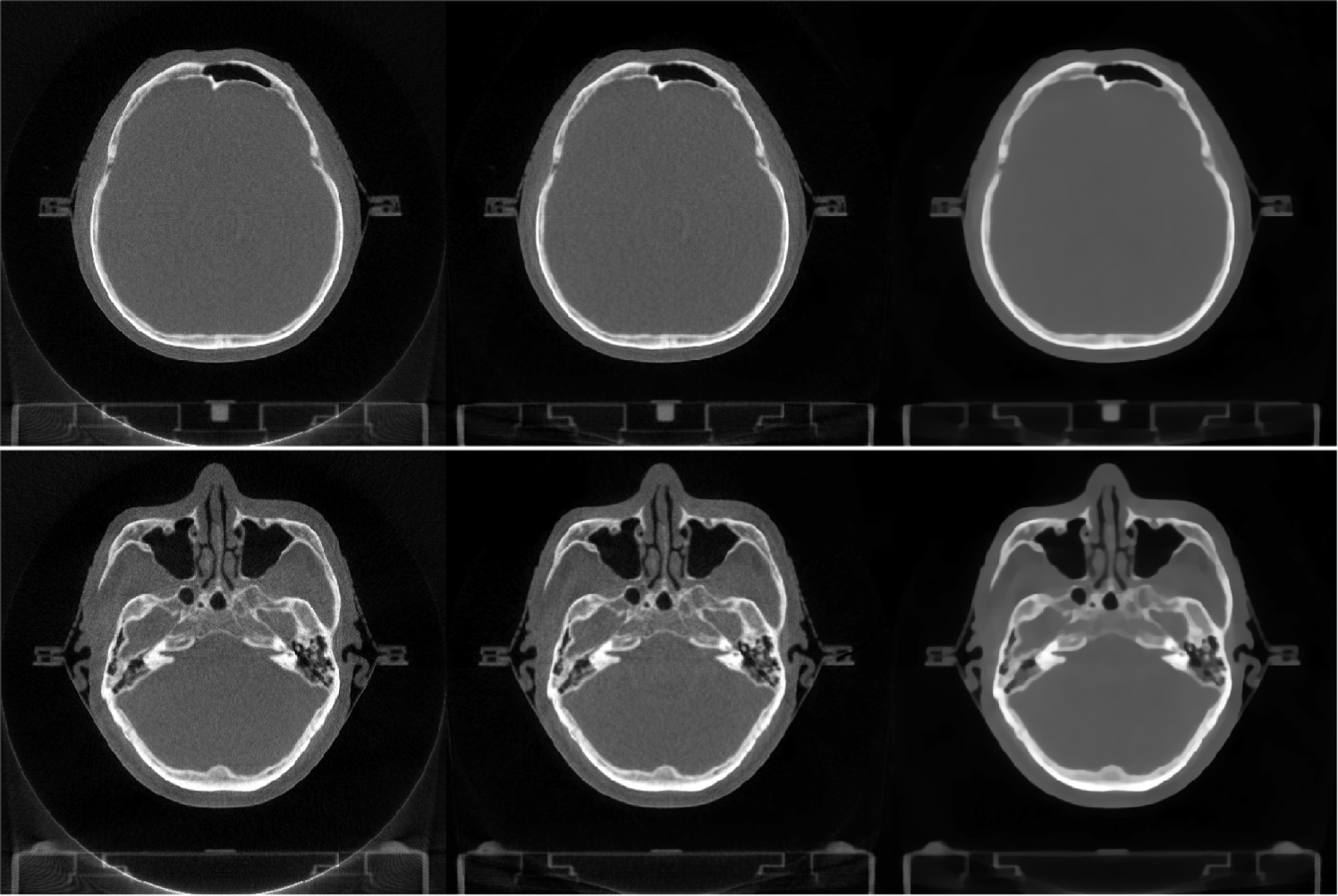
Rows are different slices; columns are FDK, OS-SART (50 iterations) and OS-ASD-POCS (50 iterations). Display window [0, 0.05].#
Thanks to Yi Liu for the data. Source: Biguri et al. (2025), TIGRE v3, figure 2 (CC BY 4.0).
Low-dose lung imaging#
Image-guided radiotherapy for lung cancer images the patient repeatedly, so every scan should use as little dose as possible. Regularised iterative algorithms remove much of the noise that FDK leaves in low-dose cone-beam CT.
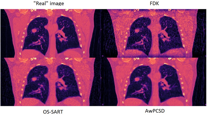
Reference image, FDK, OS-SART and AwPCSD.#
Source: Biguri et al. (2017), Physics in Medicine & Biology.
Cryo soft X-ray tomography#
Trypanosomes (single-celled parasites that cause sleeping sickness), frozen and imaged with soft X-rays at beamline B24 of the Diamond Light Source. Soft X-rays reach nanometre resolution, but the scans are very noisy and cover less than 120 degrees.


Columns: FBP, OS-SART (20 iterations) and OS-ASD-POCS (20 iterations, 20 TV iterations each). The red squares in the first image mark the zoomed areas.#
Thanks to the Diamond Light Source, especially Michele Darrow and Imanol Luengo, for sharing this dataset. Source: PhD thesis, page 101.
Synchrotron: in-situ settings#
A synchrotron scan acquired with the fast, low-dose settings of a dynamic in-situ experiment, where every algorithm has to work with noisy data.
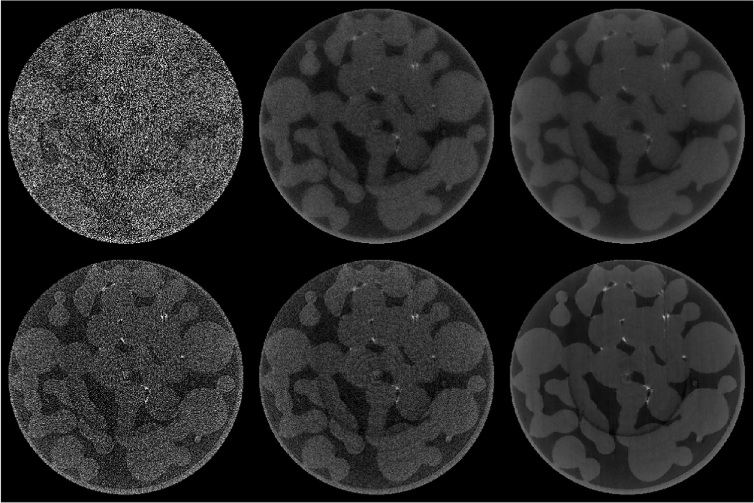
Left to right, top to bottom: FBP (17 s), LSQR with 20 iterations (249 s), FISTA with 80 iterations (1330 s), FBP with a Hamming filter (17 s), SIRT with 150 iterations (1330 s) and OS-ASD-POCS with 20 iterations (856 s).#
Source: Biguri et al. (2025), TIGRE v3, figure 3 (CC BY 4.0).
Micro-CT: SophiaBeads#
The SophiaBeads dataset was acquired for testing reconstruction methods: a 25 mm plastic tube filled with soda-lime glass beads 2.5 mm across. Here it is reconstructed from 256 projections with FDK and CGLS (15 iterations).

FDK and CGLS, 256 projections. The red line marks the profile below. Display window [0, 0.15].#

Source: PhD thesis, page 96.
Micro-CT: a very large reconstruction#
A coffee bean scanned with a shifted detector panel, reconstructed at 3340 × 3340 × 900 voxels from 2134 projections of 3780 × 900 pixels. It shows that TIGRE can reconstruct images much larger than GPU memory, even with CGLS, which needs a lot of memory.
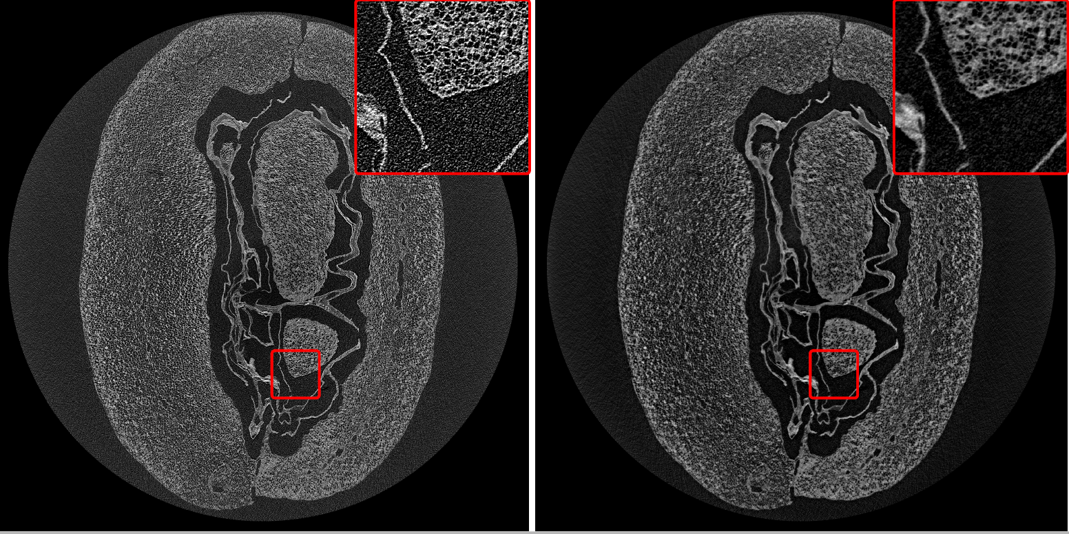
Coffee bean with FDK and CGLS (15 iterations). The image is heavily compressed; follow the links for the full-resolution originals.#


Source: Biguri et al. (2020), figure 10.
Micro-CT: a helical scan#
An Energizer AAAA battery scanned in helical mode on a diondo d5 scanner at the µ-VIS X-ray Imaging Centre, University of Southampton.
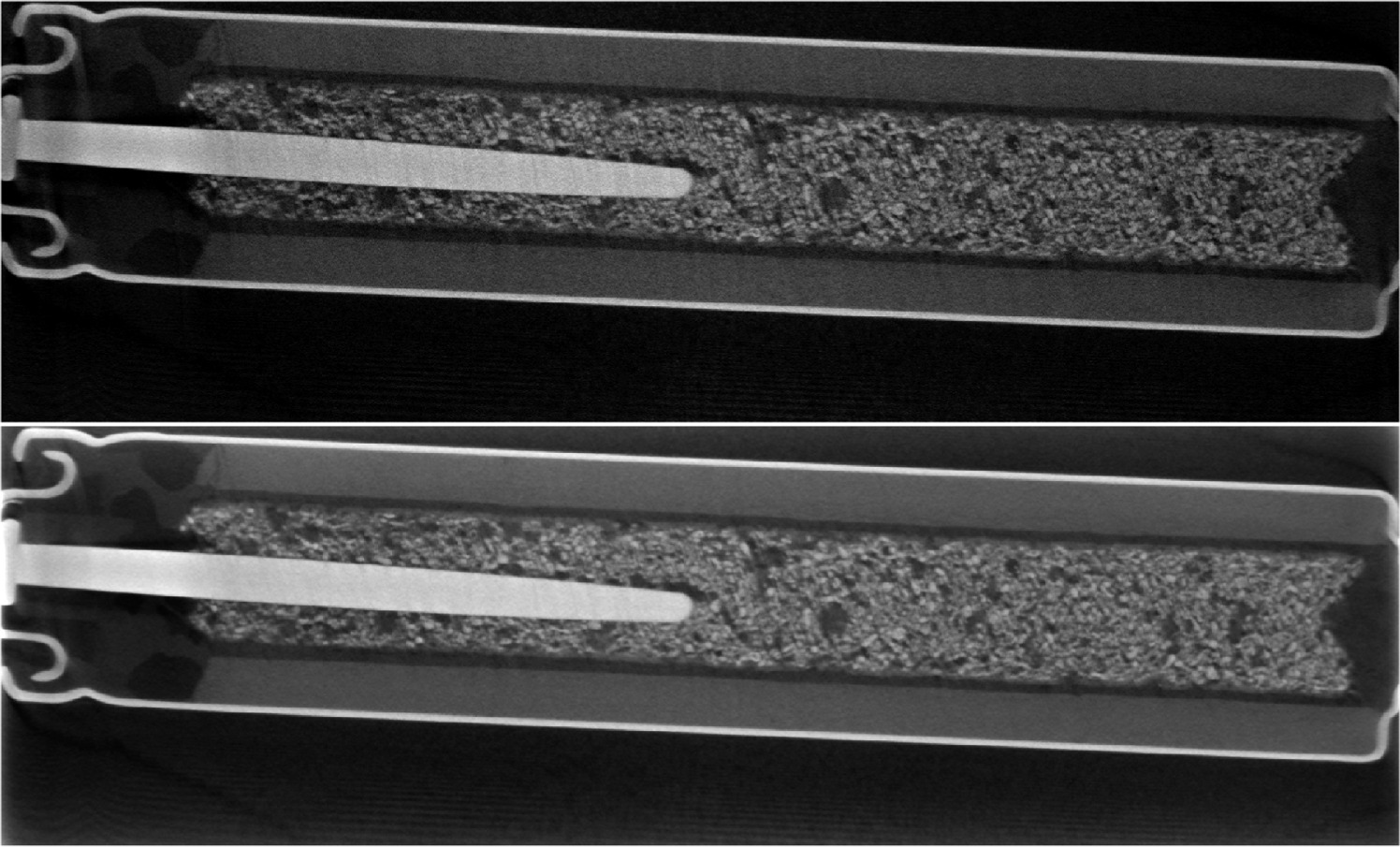
Top: FDK. Bottom: FISTA with TV regularisation (20 iterations).#
Source: Biguri et al. (2025), TIGRE v3, figure 6 (CC BY 4.0).
Neutron tomography#
A padlock imaged with neutrons at the Thailand Institute of Nuclear Technology (TINT).
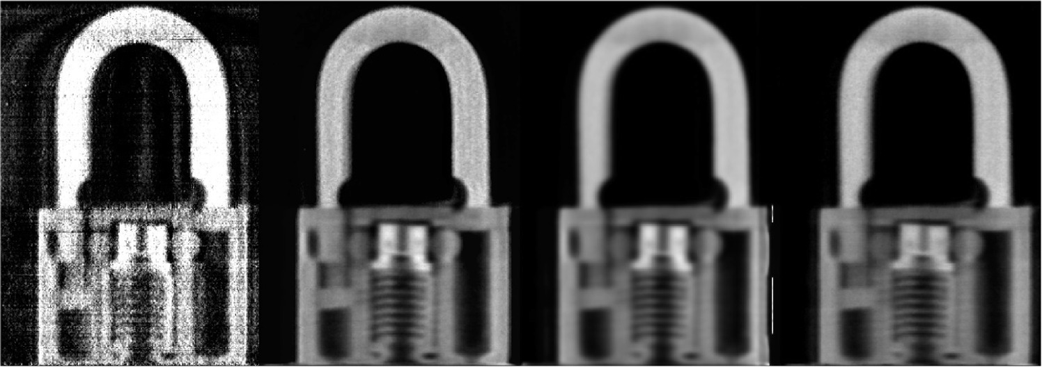
Left to right: FDK with a Shepp-Logan filter, SART, SART-TV and SIRT, the iterative ones with 100 iterations each.#
Source: Biguri et al. (2025), TIGRE v3, figure 5 (CC BY 4.0).
Proton CT#
Simulated proton CT of the high-resolution module of the Catphan phantom, reconstructed with TIGRE’s MATLAB proton CT extension (see Proton CT).
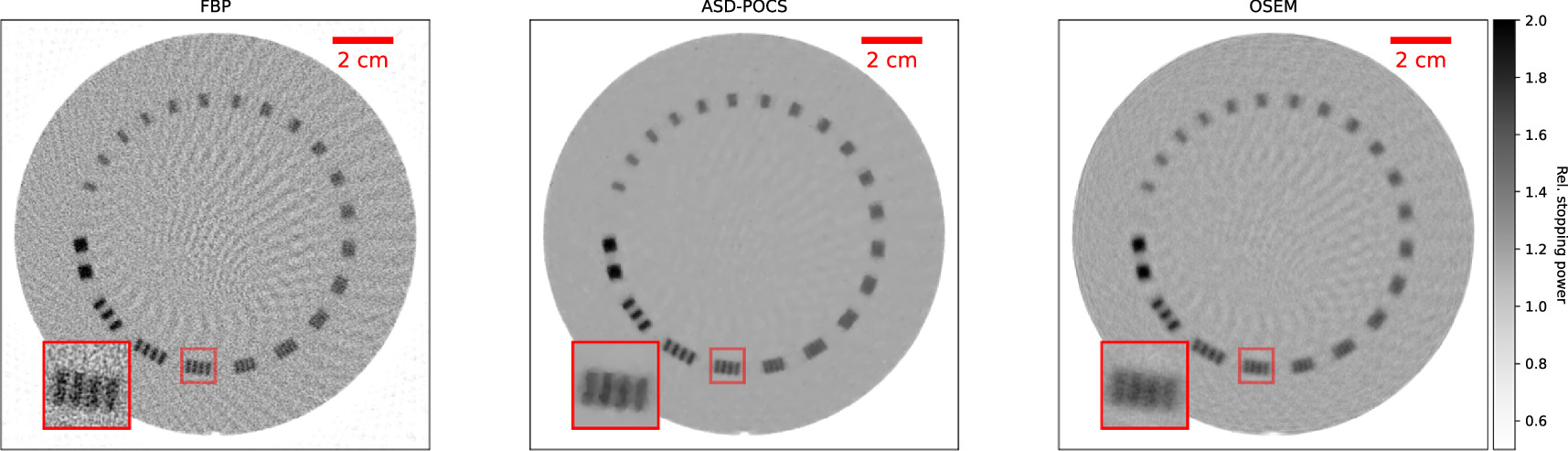
Left to right: FBP, ASD-POCS and OSEM.#
Source: Biguri et al. (2025), TIGRE v3, figure 4 (CC BY 4.0).
Designing scan trajectories#
TIGRE’s geometry can be set separately for every projection, so it can simulate scans along any source and detector path. Small dental CBCT units have a small field of view; this work uses TIGRE to design arc-based trajectories that move the scan towards chosen regions outside the standard field of view, such as the temporal bones or the spine, while using fewer projections.
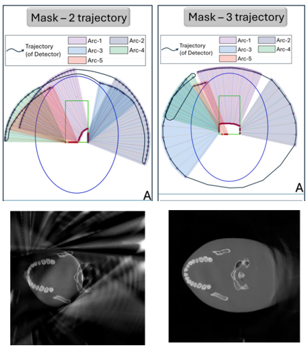
Top: trajectories designed for two regions of interest (masks 2 and 3). Bottom: reconstructions of a head from scans along them.#
Source: Islam et al. (2025), Computational and Structural Biotechnology Journal (CC BY-NC-ND 4.0).